Italian surgeon Sergio Canavero announce a project perform a human head transplant at a keynote lecture at the American Academy of Neurological and Orthopaedic Surgeons annual conference this June. He sees the procedure as being possible as soon as 2017 and believes it should be pursued as a means of saving people with, say, multi-organ cancer.
He believes the patient would be able to speak in his own voice upon waking and that walking could be achieved within a year. "If society doesn't want it, I won't do it," Canavero says. "But if people don't want it in the US or Europe, that doesn't mean it won't be done somewhere else.
Most other surgeons do not believe the procedure will be successful.
New Scientist reports that Xiao-Ping Ren of Harbin Medical University in China recently showed that it is possible to perform a basic head transplant in a mouse. Ren will attempt to replicate Canavero's protocol in the next few months in mice, and monkeys.
CNS Neuroscience and Therapeutics - Allogeneic Head and Body Reconstruction: Mouse Model
Ren's approach, pioneered in mice, involves retaining the donor brain stem and transplanting the recipient head. Our preliminary data in mice support that this allows for retention of breathing and circulatory function. Critical aspects of the current protocol include avoiding cerebral ischemia through cross-circulation (donor to recipient) and retaining the donor brain stem. Successful clinical translation of AHBR will become a milestone of medical history and potentially could save millions of people. Ren's mouse experiment confirmed a method to avoid cerebral ischemia during the surgery and solved an important part of the problem of how to accomplish long-term survival after transplantation and preservation of the donor brain stem.
Head Transplant Procedure
* The sharp severance of the cervical cords (donor's and recipient's), with its attendant minimal tissue damage
* The exploitation of the gray matter internuncial sensori-motor "highway" rebridged by sprouting connections between the two reapposed cord stumps. This could also explain the partial motor recovery in a paraplegic patient submitted to implantation of olfactory ensheathing glia and peripheral nerve bridges: A 2-mm bridge of remaining cord matter might have allowed gray matter axons to reconnect the two ends
* The bridging as per point 2 above is accelerated by electrical SCS straddling the fusion point
* The application of "fusogens/sealants": Sealants "seal" the thin layer of injured cells in the gray matter, both neuronal, glial and vascular, with little expected scarring; simultaneously they fuse a certain number of axons in the white matter.
During CSA, microsutures (mini-myelorrhaphy) will be applied along the outer rim of the apposed stumps. A cephalosomatic anastomosee will thus be kept in induced coma for 3-4 weeks following CSA to give time to the stumps to refuse (and avoid movements of the neck) and will then undergo appropriate rehabilitation in the months following the procedure.
In addition, the immunosuppressant regime that will be instituted after CSA is expected to be pro-regenerative
![]()
Figure 1: (a) Longitudinal cut along a primate spinal cord depicting the internuncial system (gray matter motor highway) and the nano-size of the proposed severance (left). The red circle on the right side of this panel is the pyramidal tract, shown in two exploded views of a sharply transected cord (middle right) and of the cord in the vertebral canal (lower middle right). (b)Visualization of the severed pyramidal tract. The uppermost image depicts a motor neuron in the cortex sending forth the axonal prolongation. Middle panel: The pyramidal tract (red) and a portion of its severed axons. Lower panel: The sharply severed axonal extensions (adapted from Laruelle 1937 and several images in the public domain)
The project for the first head transplant in man is code-named HEAVEN/GEMINI (Head Anastomosis Venture with Cord Fusion.
I covered the internet and news reactions to the 2013 discussion of technical feasibility of head transplants.
I consider the 2-13 proposed procedure in the context of organ donation and xenotransplantation.
The technical hurdles have now been cleared thanks to cell engineering. As described in his paper, the keystone to successful spinal cord linkage is the possibility to fuse the severed axons in the cord by exploiting the power of membrane fusogens/sealants. Agents exist that can reconstitute the membranes of a cut axon and animal data have accrued since 1999 that restoration of axonal function is possible. One such molecule is poly-ethylene glycol (PEG), a widely used molecule with many applications from industrial manufacturing to medicine, including as an excipient in many pharmaceutical products. Another is chitosan, a polysaccharide used in medicine and other fields.
HEAVEN capitalizes on a minimally traumatic cut of the spinal cord using an ultra-sharp blade (very different from what occurs in the setting of clinical spinal cord injury, where gross, extensive damage and scarring is observed) followed within minutes by chemofusion (GEMINI). The surgery is performed under conditions of deep hypothermia for maximal protection of the neural tissue. Moreover, and equally important, the motoneuronal pools contained in the cord grey matter remain largely untouched and can be engaged by spinal cord stimulation, a technique that has recently shown itself capable of restoring at least some motor control in spinal injured subjects.
![]()
Surgical Neurological International - HEAVEN: The head anastomosis venture Project outline for the first human head transplantation with spinal linkage (GEMINI)
* a head of a monkey was transplanted in the 1970s but the spinal cord could not be repaired at the time
* Spinal cords have been regrown in rats.
* In 2000, guinea pigs had spinal cords surgically cut and then protected with PEG chemical (like what is proposed here) and they had over 90% of spinal nerve transmission restored with a lot of mobility and function restored
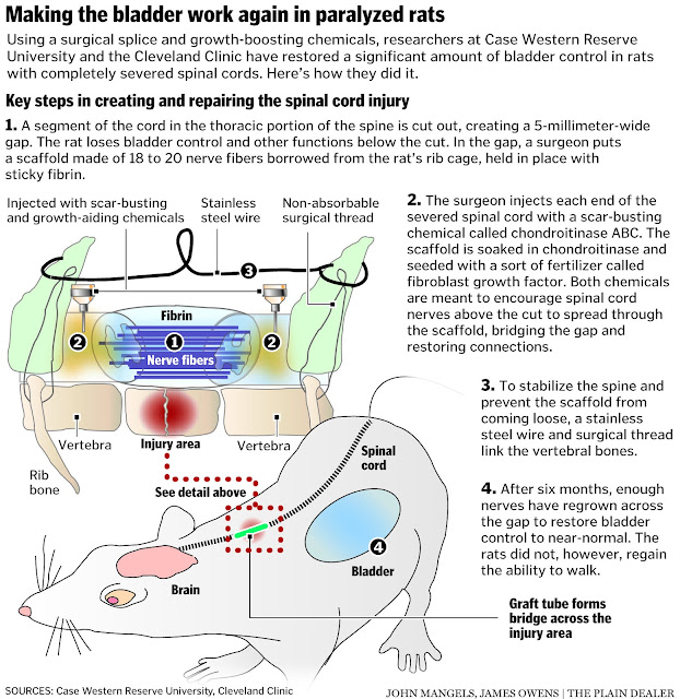
Over the last 30 years, scientists have worked to chemically encourage regrowth. Two chemicals, chondroitinase and FGF, show strong signs of doing exactly that--in rats, at least. Independently, over the past three decades, each chemical has shown some promise in restoring simple but crucial rat motor processes, like breathing, even with entirely severed spinal cords.
Two surgeons in the field figured that a combination of the chemicals might enhance the regrowth even more. The surgeons, from Case Western Reserve University and the Cleveland Clinic, began by entirely severing the spinal cords of 15 rats to ensure no independent, natural regrowth. That shut off the rats' bladder control (a nervous system process that is especially important in rats, since they urinate often and to mark their territory). The researchers then injected the two growth-stimulating chemicals into both sides of the severance, and reinforced the gap in the cord with steel wiring and surgical thread.
The Cleveland clinic has the full description of the rat spinal cord repair.
![]()
Read more »![]()
![]()
![]()
![]()
![]()
![]()
![]()
![]()
![]()
![]()
![]()
He believes the patient would be able to speak in his own voice upon waking and that walking could be achieved within a year. "If society doesn't want it, I won't do it," Canavero says. "But if people don't want it in the US or Europe, that doesn't mean it won't be done somewhere else.
Most other surgeons do not believe the procedure will be successful.
New Scientist reports that Xiao-Ping Ren of Harbin Medical University in China recently showed that it is possible to perform a basic head transplant in a mouse. Ren will attempt to replicate Canavero's protocol in the next few months in mice, and monkeys.
CNS Neuroscience and Therapeutics - Allogeneic Head and Body Reconstruction: Mouse Model
Ren's approach, pioneered in mice, involves retaining the donor brain stem and transplanting the recipient head. Our preliminary data in mice support that this allows for retention of breathing and circulatory function. Critical aspects of the current protocol include avoiding cerebral ischemia through cross-circulation (donor to recipient) and retaining the donor brain stem. Successful clinical translation of AHBR will become a milestone of medical history and potentially could save millions of people. Ren's mouse experiment confirmed a method to avoid cerebral ischemia during the surgery and solved an important part of the problem of how to accomplish long-term survival after transplantation and preservation of the donor brain stem.
Head Transplant Procedure
* The sharp severance of the cervical cords (donor's and recipient's), with its attendant minimal tissue damage
* The exploitation of the gray matter internuncial sensori-motor "highway" rebridged by sprouting connections between the two reapposed cord stumps. This could also explain the partial motor recovery in a paraplegic patient submitted to implantation of olfactory ensheathing glia and peripheral nerve bridges: A 2-mm bridge of remaining cord matter might have allowed gray matter axons to reconnect the two ends
* The bridging as per point 2 above is accelerated by electrical SCS straddling the fusion point
* The application of "fusogens/sealants": Sealants "seal" the thin layer of injured cells in the gray matter, both neuronal, glial and vascular, with little expected scarring; simultaneously they fuse a certain number of axons in the white matter.
During CSA, microsutures (mini-myelorrhaphy) will be applied along the outer rim of the apposed stumps. A cephalosomatic anastomosee will thus be kept in induced coma for 3-4 weeks following CSA to give time to the stumps to refuse (and avoid movements of the neck) and will then undergo appropriate rehabilitation in the months following the procedure.
In addition, the immunosuppressant regime that will be instituted after CSA is expected to be pro-regenerative
Figure 1: (a) Longitudinal cut along a primate spinal cord depicting the internuncial system (gray matter motor highway) and the nano-size of the proposed severance (left). The red circle on the right side of this panel is the pyramidal tract, shown in two exploded views of a sharply transected cord (middle right) and of the cord in the vertebral canal (lower middle right). (b)Visualization of the severed pyramidal tract. The uppermost image depicts a motor neuron in the cortex sending forth the axonal prolongation. Middle panel: The pyramidal tract (red) and a portion of its severed axons. Lower panel: The sharply severed axonal extensions (adapted from Laruelle 1937 and several images in the public domain)
The project for the first head transplant in man is code-named HEAVEN/GEMINI (Head Anastomosis Venture with Cord Fusion.
I covered the internet and news reactions to the 2013 discussion of technical feasibility of head transplants.
I consider the 2-13 proposed procedure in the context of organ donation and xenotransplantation.
The technical hurdles have now been cleared thanks to cell engineering. As described in his paper, the keystone to successful spinal cord linkage is the possibility to fuse the severed axons in the cord by exploiting the power of membrane fusogens/sealants. Agents exist that can reconstitute the membranes of a cut axon and animal data have accrued since 1999 that restoration of axonal function is possible. One such molecule is poly-ethylene glycol (PEG), a widely used molecule with many applications from industrial manufacturing to medicine, including as an excipient in many pharmaceutical products. Another is chitosan, a polysaccharide used in medicine and other fields.
HEAVEN capitalizes on a minimally traumatic cut of the spinal cord using an ultra-sharp blade (very different from what occurs in the setting of clinical spinal cord injury, where gross, extensive damage and scarring is observed) followed within minutes by chemofusion (GEMINI). The surgery is performed under conditions of deep hypothermia for maximal protection of the neural tissue. Moreover, and equally important, the motoneuronal pools contained in the cord grey matter remain largely untouched and can be engaged by spinal cord stimulation, a technique that has recently shown itself capable of restoring at least some motor control in spinal injured subjects.
Surgical Neurological International - HEAVEN: The head anastomosis venture Project outline for the first human head transplantation with spinal linkage (GEMINI)
* a head of a monkey was transplanted in the 1970s but the spinal cord could not be repaired at the time
* Spinal cords have been regrown in rats.
* In 2000, guinea pigs had spinal cords surgically cut and then protected with PEG chemical (like what is proposed here) and they had over 90% of spinal nerve transmission restored with a lot of mobility and function restored
Over the last 30 years, scientists have worked to chemically encourage regrowth. Two chemicals, chondroitinase and FGF, show strong signs of doing exactly that--in rats, at least. Independently, over the past three decades, each chemical has shown some promise in restoring simple but crucial rat motor processes, like breathing, even with entirely severed spinal cords.
Two surgeons in the field figured that a combination of the chemicals might enhance the regrowth even more. The surgeons, from Case Western Reserve University and the Cleveland Clinic, began by entirely severing the spinal cords of 15 rats to ensure no independent, natural regrowth. That shut off the rats' bladder control (a nervous system process that is especially important in rats, since they urinate often and to mark their territory). The researchers then injected the two growth-stimulating chemicals into both sides of the severance, and reinforced the gap in the cord with steel wiring and surgical thread.
The Cleveland clinic has the full description of the rat spinal cord repair.
Read more »


